Publicité
Les langues disponibles
Les langues disponibles
Liens rapides
A Guide to the Use of Ophthalmoscopes
Guide d'utilisation des ophtalmoscopes
Hinweise zur Verwendung des Ophthalmoskopes
Una guía para el uso de los oftalmoscopios
Una guida all'uso degli oftalmoscopi nell'esame
in the Eye Examination
pour l'examen de l'oeil
bei der Augenuntersuchung
en el examen ocular
dell'occhio
English
Français
Deutsch
Español
Italiano
Pathologies
1
8
16
24
32
40
Publicité

Sommaire des Matières pour Welch Allyn 11720
- Page 1 A Guide to the Use of Ophthalmoscopes in the Eye Examination Guide d’utilisation des ophtalmoscopes pour l’examen de l’oeil Hinweise zur Verwendung des Ophthalmoskopes bei der Augenuntersuchung Una guía para el uso de los oftalmoscopios en el examen ocular Una guida all’uso degli oftalmoscopi nell’esame dell’occhio English Français...
- Page 2 The Ophthalmoscope Transparency of the cornea, lens and vitreous humor permits the physician to directly view arteries, veins, optic nerve and the retina. Direct observation of the structures of the fundus through an ophthalmoscope may show disease of the eye itself or may reveal abnormalities indicative of disease elsewhere in the body.
- Page 3 Flourescein drops in the eye may also help detect corneal abrasions and other lesions. Other filters Welch Allyn ophthalmoscopes #11720 and #11730 are equipped with a unique sliding switch that greatly increases their versatility.
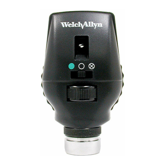
- Page 4 Red-free filter: When the switch is positioned to the left (while facing the instrument front) it will be beneath a green dot and the red-free filter will be in place. This can be used in conjunction with any aperture. The red-free filter excludes red rays from the examination field;...
- Page 5 The Eye VITREOUS HUMOR RETINA OUTSIDE LENS INSIDE LENS ANTERIOR CORNEA With the ophthalmoscope 2 inches (5cm) in front of the eye, the lenses in the rotating wheel produce clear vision at points indicated in the diagrammatic eye. The hyperopic or far-sighted eye requires more “plus” sphere for clear focus and the myopic or near-sighted eye requires “minus”...
- Page 6 A. Macula J. Cornea B. Vitreous humor K. Ciliary Body C. Sclera L. Zonule (Suspensory Ligament) D. Choroid M. Conjunctiva E. Retina N. Lens F. Ora Serrata O. Hyaloid canal G. Canal of Schlemm P. Central retinal vein H. Anterior chamber Q.
- Page 7 How to Conduct an Ophthalmologic Examination Position the ophthalmoscope about 6 inches (15cm) in front and 25° to the right side of the patient. (Step 5) In order to conduct a successful examination of the fundus, the examining room should be either semi-darkened or completely darkened.
- Page 8 6. Rest the left hand on the patient’s forehead and hold the upper lid of the eye near the eyelashes with the thumb. While the patient holds his fixation on the specified object, keep the “reflex” in view and slowly move toward the patient.
- Page 9 8. To examine the extreme periphery, instruct the patient to: a) look up for examination of the superior retina b) look down for examination of the inferior retina c) look temporally for examination of the temporal retina d) look nasally for examination of the nasal retina. This routine will reveal almost any abnormality that occurs in the fundus.
- Page 10 L’ophtalmoscope La transparence de la cornée, du cristallin et de l’humeur vitrée permet au praticien d’examiner directement les artères, les veines, le nerf optique et la rétine. L’observation directe des structures du fond de l’œil à travers un ophtalmoscope peut révéler soit des troubles de l’œil même, soit des anomalies caractéristiques de troubles présents dans d’autres régions du corps.
-
Page 11: Autres Filtres
L’administration de gouttes fluorescentes dans l’œil peut également permettre de détecter les abrasions de la cornée et d’autres lésions. Autres filtres Les ophtalmoscopes Welch Allyn n° 11720 et 11730 sont munis d’un sélecteur coulissant exclusif qui accroît considérablement leur polyvalence. -
Page 12: Autres Emplois De L'ophtalmoscope
Filtre sans rayons rouges : Lorsqu’on place le sélecteur à gauche (en faisant face à l’avant de l’instrument), il est situé au-dessous d’un point vert ; le filtre sans rayons rouges est alors en place. Ce filtre peut être utilisé avec n’importe quelle ouverture. Il exclut les rayons rouges du champ d’examen, ce qui assure des conditions supérieures à... - Page 13 L’œil HUMEUR VITRÉE CRISTALLIN RÉTINE EXTÉRIEUR CRISTALLIN INTÉRIEUR CORNÉE ANTÉRIEURE Lorsque l’ophtalmoscope est à 5 cm (2 po) de l’œil, les lentilles du sélecteur rotatif permettent de voir clairement les points indiqués sur le schéma de l’œil. L’œil hypermétrope ou presbyte requiert des lentilles « positives » pour une pour une mise au point claire tandis que l’œil myope requiert des lentilles «...
- Page 14 A. Macula J. Cornée B. Humeur vitrée K. Corps ciliaire C. Sclérotique L. Zonule (ligament suspenseur) D. Choroïde M. Conjonctive E. Rétine N. Cristallin F. Ora serrata O. Canal hyaloïdien G. Canal de Schlemm P. Veine rétinienne centrale H. Chambre antérieure Q.
-
Page 15: Comment Effectuer Un Examen Ophtalmoscopique
Comment effectuer un examen ophtalmoscopique Placer l’ophtalmoscope à environ 15 cm (6 po) du patient, à un angle de 25° à sa droite (étape 5). Pour effectuer un bon examen du fond de l’œil, obscurcir la salle d’examen à demi ou totalement. Il est préférable de dilater la pupille s’il n’y a pas de contre-indication... - Page 16 6. Poser la main gauche sur le front du patient et tenir avec le pouce sa paupière supérieure, à proximité des cils. Pendant que le patient fixe l’objet, garder le « réflexe » en vue et s’approcher lentement du patient. La papille optique devrait lorsqu’on se trouve à...
- Page 17 8. Pour examiner la périphérie, demander au patient : a) de regarder en haut pour l’examen de la rétine supérieure b) de regarder en bas pour l’examen de la rétine inférieure c) de regarder de côté pour l’examen de la rétine temporale d) de regarder son nez pour l’examen de la rétine nasale.
- Page 18 Das Ophthalmoskop Die Transparenz der Kornea, der Augenlinse und des Glaskörpers gestatten es dem Arzt, Arterien, Venen, Sehnerv und Retina direkt einzusehen. Durch die direkte Betrachtung der Fundusstrukturen mit einem Ophthalmoskop können Erkrankungen des Auges selbst oder Abnormalitäten, die auf Erkrankungen anderer Körperteile zurückzuführen sind, festgestellt werden.
- Page 19 Gefäßprobleme, z.B. Gefäßblutungen oder eine erhöhte Gefäßwandddurchlässigkeit, erkannt werden. Durch Einträufeln von Fluoreszeinlösung in das Auge sind Hornhauterosionen und ander Läsionen ebenfalls erkennbar. Weitere Filter Die Welch Allyn Ophthalmoskope Nr. 11720 und 11730 sind mit einer zusätzlichen Blendenvorschaltung ausgerüstet, die ihre Vielseitigkeit noch enorm erhöht.
- Page 20 Rotfrei-Filter: Steht der Schalter in der linken Position (von der Patientenseite aus gesehen) unter einem grünen Punkt, so ist der Rotfrei-Filter eingeschaltet. Dieser Filter kann nun mit einer beliebigen Blende kombiniert werden. Dies ist gegenüber einer Untersuchung mit Weißlicht von Vorteil, da kleine Gefäßveränderungen, winzige Netzhautblutungen, diffuse Exsudate und ungewöhnliche Veränderungen in der Macula leichter festzustellen sind.
- Page 21 Das Auge GLASKÖRPER ÄUSSERER RETINA LINSENBEREICH INNERER LINSENBEREICH VORDERER KORNEABEREICH Bei einem Abstand des Ophthalmoskops von 5 cm vor dem Auge sind über die entsprechenden Linsen in der Linsenwahlscheibe von den in obigem Diagramm angezeigten Augenbereichen scharfe Darstellungen möglich. Bei Weitsichtigkeit (Hyperopie) sind für eine scharfe Darstellung Linsen mit positiven Werten und bei Kurzsichtigkeit (Myopie) Linsen mit negativen Werten erforderlich.
- Page 22 A. Macula J. Kornea K. Ziliarkörper B. Glaskörper L. Zonula Fasern C. Lederhaut (Sklera) M. Bindehaut (Konjunktiva) D. Aderhaut (Choroidea) N. Linse O. Canalis hyaloideus E. Netzhaut (Retina) P. Zentrale Retinavene F. Ora Serrata Q. Sehnerv G. Schlemm´scher Kanal R. Zentrale Retinaarterie H.
- Page 23 Durchführung einer ophthalmologischen Untersuchung Das Ophthalmoskop etwa 15 cm vor und 25° rechts vom Patienten ausrichten (siehe Schritt 5). Für eine erfolgreiche Untersuchung des Fundus sollte das Behandlungszimmer nur schwach beleuchtet oder ganz abgedunkelt sein. Liegen keine Kontraindikationen vor, werden die Pupillen erweitert, jedoch sind auch bei nicht erweiterten Pupillen viele wertvolle...
- Page 24 6. Sich dem Auge des Patienten auf ca. 5 cm nähern. Dabei kann die linke Hand auf dem Kopf ruhen und der linke Daumen das Oberlid hochhalten. Während der Patient den Blick weiter auf das entfernte Objekt fixiert, behalten Sie den „Reflex“ im Auge und bewegen sich langsam auf den Patienten zu.
- Page 25 8. Zur Untersuchung der Peripherie der Netzhaut fordern Sie den Patienten auf: a) nach oben zu sehen für die Untersuchung der oberen Retina b) nach unten zu sehen für die Untersuchung der unteren Retina c) auf die Seite zu sehen für die Untersuchung der temporalen Retina d) zur Nase zu sehen für die Untersuchung der nasalen Retina.
- Page 26 El oftalmoscopio La transparencia de la córnea, el cristalino y el humor vítreo permiten al médico ver directamente las arterias, las venas, el nervio óptico y la retina. La observación directa de las estructuras del fondo mediante un oftalmoscopio puede mostrar enfermedades del ojo mismo o puede revelar anormalidades indicadoras de enfermedades en otras partes del cuerpo.
- Page 27 Otros filtros Los oftalmoscopios No. 11720 y No. 11730 de Welch Allyn están equipados con un interruptor deslizante único que aumenta en gran medida su versatilidad.
- Page 28 Filtro sin rojo: Cuando el interruptor está ubicado hacia la izquierda (mirando la parte frontal del instrumento) estará debajo de un punto verde y el filtro sin rojo estará en su lugar. Esto se puede utilizar en conjunto con cualquier apertura. El filtro sin rojo excluye los rayos rojos del campo de examen;...
- Page 29 El ojo HUMOR VÍTREO CRISTALINO RETINA EXTERIOR CRISTALINO INTERIOR CORNEA FRONTAL Con el oftalmoscopio a 5 cm (2 pulgadas) de la parte frontal del ojo, las lentes de la rueda giratoria producen una visión clara en los puntos indicados en el diagrama del ojo. El ojo hiperópico o hipermetrópico requiere mayor esfera “más”...
- Page 30 A. Mácula J. Córnea B. Humor vítreo K. Cuerpo ciliar C. Esclerótica L. Zónula (ligamento suspensor) D. Coroides M. Conjuntiva E. Retina N. Cristalino F. Ora serrata O. Canal Hialoideo G. Canal de Schlemm P. Vena retinal central H. Cámara anterior Q.
- Page 31 Cómo realizar un examen oftalmológico Coloque el oftalmoscopio a unos 15 cm (6 pulgadas) al frente y 25° hacia la derecha del paciente (Paso 5). A fin de efectuar un examen satisfactorio del fondo, la sala de examen debe estar semioscura o completamente oscura.
- Page 32 6. Apoye la mano izquierda sobre la frente del paciente y sostenga el párpado superior del ojo cerca de las pestañas con el pulgar. Mientras el paciente sostiene fija su mirada en el objeto especificado, mantenga el “reflejo” a la vista y lentamente muévase hacia el paciente.
- Page 33 8. Para examinar la periferia extrema pida al paciente que: a) mire hacia arriba para examinar la retina superior b) mire hacia abajo para examinar la retina inferior c) mire en dirección temporal para examinar la retina temporal d) mire en dirección nasal para examinar la retina nasal Esta rutina revelará...
- Page 34 L’oftalmoscopio La trasparenza della cornea del cristallino e dell’humor vitreo, permette di vedere direttamente le arterie le vene, il nervo ottico e la retina. L’osservazione diretta delle strutture del fondo attraverso un oftalmoscopio può evidenziare non solo malattie sello stesso occhio, ma anche rivelare alcune anormalità indicative di patologie di altre parti dell’organismo.
- Page 35 Le gocce di fluorosceina nell’occhio possono inoltre essere di ausilio nella rivelazione di abrasioni alla cornea o di altre lesioni. Altri filtri I modelli 11720 e 11730 degli oftalmoscopi Welch Allyn sono muniti di un originale selettore di filtri che ne aumenta considerevolmente la versatilità.
- Page 36 Filtro rosso privo: Quando l’interruttore si trova spostato a sinistra osservando la parte anteriore dello strumento, questo apparirà in corrispondenza di un punto verde, a indicare che il filtro rosso privo è in posizione. Questa impostazione può essere usata unitamente a una qualsiasi apertura, dato che detto filtro esclude le radiazioni rosse dal campo in esame.
- Page 37 L’occhio HUMOR VITREO ESTERNO DEL RETINA CRISTALLINO INTERNO DEL CRISTALLINO CORNEA ANTERIORE Mentre si tiene l’oftalmoscopio a 5 cm dall’occhio, le lenti presenti nella ruota di selezione permettono una chiara visione dei punti indicati nel diagramma dell’occhio. L’occhio presbite, o che vede da lontano, richiede una maggiore curvatura per la messa a fuoco, l’occhio miope, o che vede da vicino, richiede una minore curvatura.
- Page 38 A. Macula J. Cornea B. Humor vitreo K. Corpo ciliare C. Sclera L. Zonula (legamento sospensorio) D. Coroide M. Congiuntiva E. Retina N. Cristallino F. Ora serrata O. Canale ialoide G. Canale di Schlemm P. Vena retinica centrale H. Camera anteriore Q.
- Page 39 Conduzione di un esame oftalmologico Posizione dell’oftalmoscopio a circa 15 cm ed a 25° sulla parte destra del paziente (Punto 5). Al fine di fare un buon esame del fondo, il locale dovrebbe essere semi buio o completamente oscurato. È preferibile dilatare la pupilla quando non sussistono controindicazioni, anche se...
- Page 40 6. Tenere la mano sinistra sulla fronte del paziente e sollevare con il pollice la parte superiore della palpebra vicino alle sopracciglia. Mentre il paziente continua a fissare uno specifico oggetto, osservare il riflesso e lentamente avvicinarsi al paziente. Il disco ottico dovrebbe apparire quando si è...
- Page 41 8. Per esaminare le zone periferiche estreme, invitare il paziente a: a) guardare in alto per esaminare la retina superiore; b) guardare in basso per esaminare la retina inferiore; c) guardare in direzione delle tempie per esaminare la retina temporale; d) guardare in direzione del naso per esaminare la retina nasale.
- Page 42 Common Pathology of the Eye Häufige pathologische Befunde am Augenhintergrund/ Pathologie courante de l’œil/Patologías comunes del ojo/ Patologie comuni dell’occhio Normal fundus Disc: Outline clear; physiological cup is pale area centrally Retina: Normal red/orange color, macula is dark avascular area temporally Vessels: Arterial —...
- Page 43 Fondo normale Disco: Contorno nitido, la coppa fisiologica è un’area pallida verso il centro. Retina: Normale se di colore rosso/arancio; la macula corrisponde all’area avascolare scura nella zona temporale. Vasi: Rapporto arterie/vene di 2 a 3; le arterie appaiono di un rosso brillante, le vene di colore leggermente purpureo.
- Page 44 Proliferative diabetic retinopathy Disc: Net of new vessels growing on disc surface Retina: Numerous hemorrhages, new vessels at superior disc margin Vessels: Dilated retinal veins Rétinopathie diabétique proliférante Papille : Apparition d’un réseau de nouveaux vaisseaux à la surface de la papille Rétine : Nombreuses hémorragies, nouveaux vaisseaux sur le bord...
- Page 45 Papilledema Disc: Elevated, edematous disc, blurred disc margins; vessels engorged Retina: Flame retinal hemorrhage close to disc Vessels: Engorged tortuous veins Œdème papillaire Papille : Papille surélevée et œdémateuse au bord flou ; vaisseaux engorgés Rétine : Hémorragie rétinienne en flammèches près de la papille Vaisseaux : Veines sinueuses et engorgées Papillenödem Papille:...
- Page 46 Benign choroidal nevus Retina: Slate gray, flat lesion under retina; several drusen overlying nevus Vessels: Normal Naevus choroïdien bénin Rétine : Lésion plate de couleur gris ardoise sous la rétine ; plusieurs corps colloïdes sus-jacents Vaisseaux : Normaux Gutartiger Choroid-Nävus Retina: Schiefergrau, flache Läsion unter der Retina;...
- Page 47 Retinal detachment Disc: Normal Retina: Gray elevation in temporal area with folds in detached section Vessels: Tortuous and elevated over detached retina Décollement de la rétine Papille : Normale Rétine : Soulèvement de couleur grise dans la région temporale avec replis dans la section décollée Vaisseaux : Sinueux et surélevés sur la rétine décollée Netzhautablösung...
- Page 48 Welch Allyn, Inc. 4341 State Street Road P.O. Box 220 Skaneateles Falls, NY 13153-0220 U.S.A. Phone: (800) 535-6663 FAX: (315) 685-3361 (315) 685-4560 PM117072-01 Rev. B Printed in U.S.A.


